Quality assurance measurements ensure that the machine generates just as much X-ray as needed to keep radiation exposure as low as possible to achieve good quality imaging and avoid repeated procedures. Quality assurance testing is done for the safety of both patients and staff. It also enables predictive maintenance, and can help extend the lifetime of the machine.
Dentists use radiography to find malignant masses, bone loss, cavities, and bone density changes e.g. caries.
Dental X-ray examinations can be intraoral or panoramic:
- Intraoral examinations are done from within the mouth. Radiation penetrates oral structures before striking the film or digital receptor, placed between the patient´s teeth.
- Panoramic dental radiography involves complete scanning of the upper and lower jaw enabling a 3D view. The X-ray tube rotates, with the detector opposite to it, in a semicircle around the head, starting at one side and ending on the other.
The X-ray radiation dose for a dental patient is typically very small, equivalent to a few days of environmental background radiation. Panoramic X-ray exposure can be slightly higher than for intraoral procedures.
Teeth appear lighter because less radiation penetrates them to reach the detector. Dental caries appears darker since X-rays more easily penetrate less dense structures. Dental restorations (fillings, crowns) may appear lighter or darker, depending on material density.
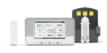
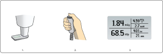
Measure with RaySafe X2 Solo Dent or X2 R/F sensor
Turn on Base Unit, and connect the sensor.
- Position the sensor or the panoramic holder with the X2 sensor attached. Make sure it is centered, and that the whole sensor is within the direct beam.
- Expose.
- Read result from Base Unit. All parameters listed above for this sensor type are gained within the same exposure.
For comprehensive analysis use the RaySafe View software.


Measure with RaySafe ThinX sensor
- Position the RaySafe ThinX on a flat surface with the collimator of the X-ray machine close to the sensor area.
- Expose.
- Read result from ThinX.
Watch how-to-video:
RaySafe X2 on dental panoramic machines
White paper:
Why it is important to perform quality assurance testing for diagnostic X-ray
Case Study:
Public Dental Care Benefits from Facilitated X-ray QA-testing
Product Catalog:
X-ray Test Equipment Product Catalog
Brochures & Flyer:
RaySafe X2 Solo Dent Brochure
RaySafe X2 Solo Specification Brochure
RaySafe ThinX Brochure
Flyer: Why do quality assurance testing of X-ray equipment?
Manuals:
RaySafe X2 Solo User Manual
RaySafe ThinX Intra User Manual
RaySafe ThinX Rad User Manual
Suitable products for measurements on dental X-ray machines
Contact us for more information.
ContactThe history of dental X-ray
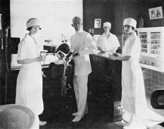
In January 1896, German dentist Otto Walkhoff made the first radiograph of his own teeth. Exposure time was 25 minutes and image quality was rather poor! Image quality was rapidly improved by many early pioneers, and in July 1896, Charles Edmund Kells, did the first dental X-ray on a patient. However, it took some time until dental X-ray was commercialized and widespread. In 1905, the first dental machines were manufactured in Germany by what is now Siemens.
Panoramic radiographic is a specific X-ray modality, designed specifically for dental applications in order to get a single radiograph with all teeth in both the upper and lower jaws. The first panoramic unit was invented by Yrjö Paatero, a Finnish dentist. Yrjö teamed up with Timo Nieminen. Together they developed the panoramic technology further and in the 1960´s, a cooperation with Siemens was established. Many important improvements have been made ever since, like reduction of dose which in the early days was considerable higher compared to intraoral procedures.
The era of digital receptors started in 1987. Throughout the years, improvements have been made regarding resolution, dynamic range, and signal-to-noise ratio. When semiconductor receptors were introduced in 1998, dental X-ray was revolutionized, in regards to image quality, exposure time, dose reduction, and time savings.
